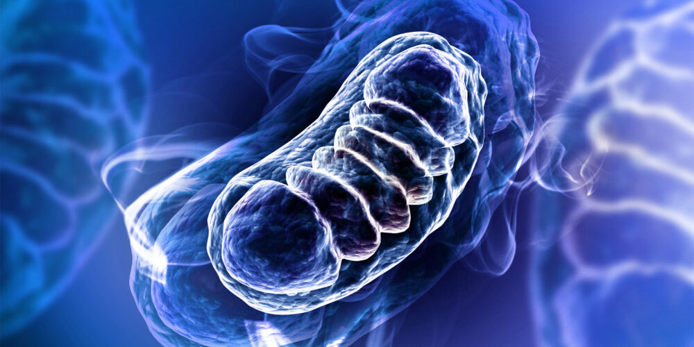
Mitochondria are known as the powerhouses of the cell and have an important role in energy production, as well as involvement in apoptosis, calcium signalling, regulation of cellular metabolism and generation of haem and steroids.
Mitochondria have a distinct structure with an outer and inner membrane. The inner membrane is highly convoluted to form folds known as cristae. Most of the cell’s supply of ATP, which is its primary energy source, is formed on these cristae. Five complexes (complex I, II, III, IV and V) are located in the cristae and these complexes are involved in oxidative phosphorylation and ATP production.
The number of mitochondria in a cell varies depending on tissue type and its function. For example, high energy requiring organs such as skeletal muscle, the heart, the brain and the liver contain numerous mitochondria, unlike mature red blood cells which have none.
Mitochondrial Toxicity Services
The mitochondria are a common target for drug-induced toxicity. We offer a number of different methods for assessing in vitro mitochondrial toxicity. These include:
- The functional mitochondrial toxicity assay detects effects of test article on oxygen consumption rate (OCR) and extracellular acidification rate (ECAR) using the Seahorse XF Technology to assess cellular metabolism and mitochondrial function. A mitochondrial stress test is used to understand cellular bioenergetics and the mechanism of mitochondrial toxicity.
- Permeabilisation of the cell membrane leaves the mitochondrial membrane intact which allows study of mitochondrial function without the need to isolate mitochondria. By using complex-specific substrates and inhibitors, it allows the identification of the individual complexes which are involved in mitochondrial toxicity. Find out more about our mitochondrial respiratory complex assay.
- Mitochondrial biogenesis can be detected using high content imaging to determine the ratio of COX-1 (mtDNA-encoded protein) to SDH-A (nDNA-encoded protein).
- Many cell lines developed for in vitro use are metabolically adapted for growth under hypoxic and anaerobic conditions in high glucose media and derive most of their energy from glycolysis rather than mitochondrial oxidative phosphorylation – a process known as the Crabtree effect. Under these conditions, mitochondrial toxicants have reduced susceptibility. By replacing glucose with galactose in the cell media, it circumvents the Crabtree effect, which increases the reliance of the cells on mitochondrial oxidative phosphorylation to obtain ATP. By investigating the toxic effects of different drugs in the glucose and galactose media, the mitochondrial Glu/Gal assay is an ideal screening assay to detect effects on mitochondrial function and identify if this is a primary effect or secondary to other cytotoxic mechanisms
- Using high content imaging, mitochondrial mass and mitochondrial membrane potential can be detected using specific cellular dyes. Find out more about our HCS mitochondrial assay.

