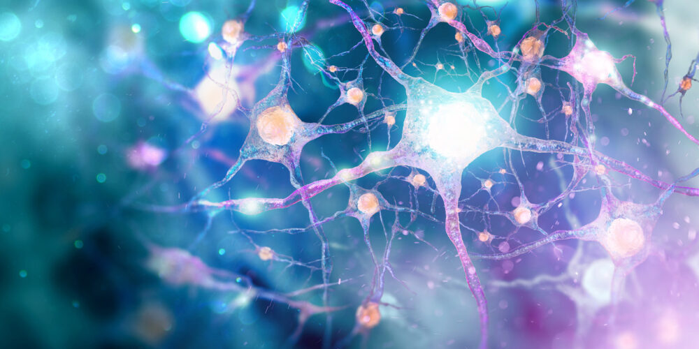
Detect neurotoxicity of novel therapeutics with enhanced in vivo relevance using Cyprotex’s 3D brain microtissue combined with high content imaging (HCI) end points.
Cyprotex delivers consistent, high quality data with the flexibility to adapt protocols based on specific customer requirements.
Introduction
- The prevalence of adverse neurotoxic reactions of the brain in response to drugs or environmental hazards continues to prompt the development of novel cell-based assays for accurate neurotoxicity prediction1.
- In vitro three-dimensional cell cultures allow better recapitulation of the complex in vivo microenvironment than traditional 2D monolayer models2.
- 3D models also permit long-term compound exposures allowing a closer replication of clinical dosing strategies3.
- Mitochondrial dysfunction and calcium homeostasis4 are commonly observed responses to toxic compounds and are implicated in neurotoxicity.
- Confocal high content imaging (HCl) allows the simultaneous detection of multiple cell health parameters within a 3D microtissue structure in combination with a measure of cellular ATP content.
Protocol
3D Neurotoxicity Assay Protocol
Data
Data from Cyprotex's 3D Neurotoxicity Assay
References
1) Wilson MS et al., (2014) Multiparametric high content analysis for assessment of neurotoxicity in differentiated neuronal cell lines and human embryonic stem cell-derived neurons. Neurotoxicology 42; 33-48
2) Anderl JL et al., (2009) A neuronal and astrocyte co-culture assay for high content analysis of neurotoxicity. J Vis Exp 27; e1173
3) Pamies D et al., (2017) A human brain microphysiological system derived from induced pluripotent stem cells to study neurological diseases and toxicity. ALTEX 34(3); 362-376
4) Guo GW & Liang YX (2001) Aluminium-induced apoptosis in cultured astrocytes and its effect on calcium homeostasis. Brain Res 888; 221-226

